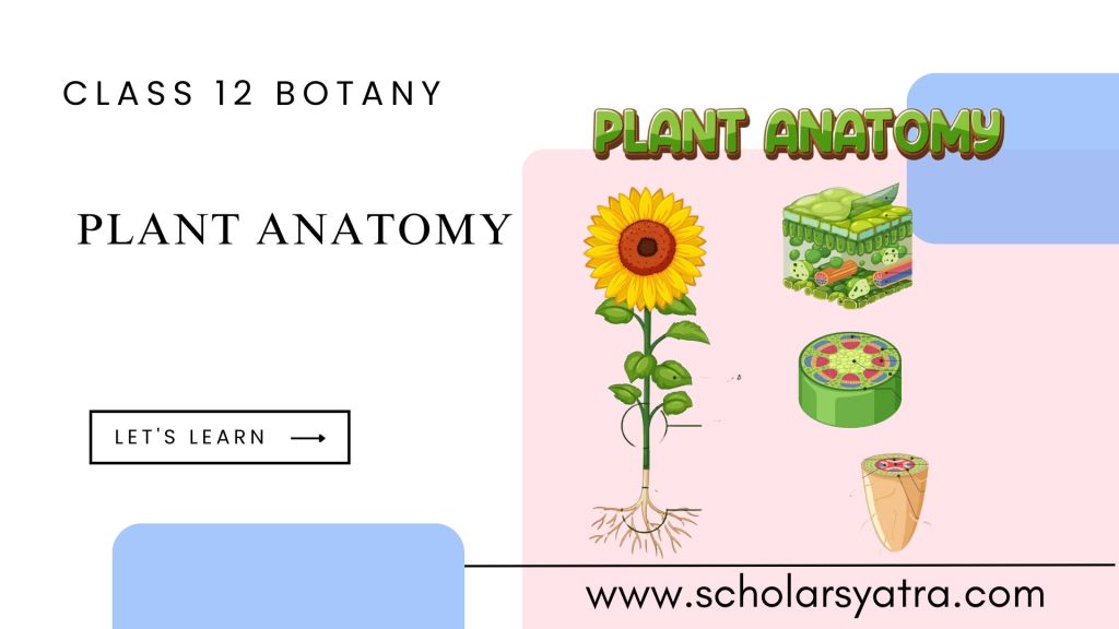
Plant anatomy is a fundamental aspect of botany that helps us understand the internal structure of plants. Students and researchers must have a comprehensive understanding of plant tissues, including their types and roles, as well as the anatomy of different parts of plants such as roots, stems, and leaves. This guide will cover these topics in detail, offering insights into plant tissues, the specific anatomy of dicot and monocot plants, and an explanation of secondary growth in dicot stems.
Table of Contents
ToggleConcept of Tissues in Plant Anatomy
Plant tissues are groups of cells that work together to perform specific functions. Tissues are the basic part of plant anatomy. These tissues can be broadly categorized into two main types: meristematic tissues and permanent tissues. Understanding these classifications is essential for defining plant structure and function.
Types of Plant Tissues
Mainly there are two types of plant tissues: Namely Meristematic tissues, and Permanent Tissues
-
Meristematic Tissues
Meristematic tissues are composed of actively dividing cells that contribute to the growth of the plant. These cells are undifferentiated, allowing them to divide and form new tissues continuously.
-
Characteristics of Meristematic Tissues:
- Cells are small, with a dense cytoplasm and a prominent nucleus.
- Lack of vacuoles and possess thin primary cell walls.
- High metabolic activity to support rapid cell division.
-
General Types of Meristems:
- Apical Meristem: Found at the tips of roots and shoots, responsible for primary growth and elongation.
- Lateral Meristem: Found along the sides of stems and roots, contributing to secondary growth (e.g., vascular cambium and cork cambium).
- Intercalary Meristem: Present at the base of leaves or internodes, facilitating regrowth and elongation.
Further Meristematic tissues are classified based on origin, position, and function.
-
Meristematic Tissue Based on Origin:
- Primary Meristem: These are derived directly from embryonic cells and contribute to the primary growth of the plant. Examples include apical meristems found at the tips of roots and shoots.
- Secondary Meristem: These originate from previously differentiated cells that regain their capacity to divide. They are responsible for secondary growth, such as the vascular cambium and cork cambium in woody plants.
-
Meristematic Tissue Based on Position:
- Apical Meristem: Located at the tips of roots and shoots, responsible for the increase in length (primary growth) of the plant.
- Lateral Meristem: Found along the sides of stems and roots, contributing to an increase in thickness or girth (secondary growth). Examples include the vascular cambium and cork cambium.
- Intercalary Meristem: Present at the base of leaves or internodes, particularly in grasses and other monocots. It aids in regrowth and lengthening of plant parts.
-
Meristematic Tissue Based on Function:
- Protoderm: Forms the epidermal tissue system and is involved in producing the outer layer of cells.
- Procambium: Gives rise to the vascular tissues (xylem and phloem) for the transport of water, nutrients, and food.
- Ground Meristem: Develops into the ground tissue system, which includes parenchyma, collenchyma, and sclerenchyma, contributing to support, storage, and photosynthesis.
2. Permanent Tissues
Permanent tissues are derived from meristematic tissues and have lost their ability to divide. They are specialized for various functions and can be classified as simple or complex.
Simple Permanent Tissues:
These tissues are made up of only one type of cell that is structurally and functionally similar. They perform basic supportive and protective roles in the plant.
- Parenchyma:
- Structure: Composed of living cells with thin, flexible cell walls and a large central vacuole. The cells can be loosely or densely packed with intercellular spaces.
- Function: Functions in storage, photosynthesis (in chlorenchyma with chloroplasts), and basic metabolic activities. Parenchyma cells in aquatic plants may have air spaces (aerenchyma) for buoyancy.
- Collenchyma:
- Structure: Consists of living cells with unevenly thickened cell walls, mainly at the corners, providing flexibility.
- Function: Provides mechanical support and elasticity to growing parts of the plant, allowing them to bend without breaking.
- Sclerenchyma:
- Structure: Composed of dead cells with thick, lignified cell walls. These cells lack intercellular spaces and come in two forms: fibers (long and thin) and sclereids (varied shapes, often shorter).
- Function: Provides rigidity and strength to the plant, contributing to its structural support. Sclerenchyma cells are found in mature parts of the plant such as stems, bark, and seed coats.
Complex Permanent Tissues:
Complex permanent tissues are composed of more than one type of cell, working together as a unit to perform specific functions, primarily in transport and support.
- Xylem:
- Structure: Made up of different types of cells, including tracheids, vessels, xylem parenchyma, and xylem fibers. Tracheids and vessels are dead at maturity and have thick, lignified walls for water conduction.
- Function: Conducts water and minerals from roots to different parts of the plant and provides structural support.
- Phloem:
- Structure: Composed of various cell types such as sieve tube elements, companion cells, phloem fibers, and phloem parenchyma. Unlike xylem, most of the phloem cells are living at maturity.
- Function: Transports food and nutrients, primarily sugars produced during photosynthesis, from the leaves to other parts of the plant.
Anatomy of Dicot and Monocot Roots
a. Dicot Root Anatomy
- Epidermis: Outer protective layer.
- Cortex: Composed of parenchyma cells that store food and transport water and minerals.
- Endodermis: The innermost layer of the cortex, regulates water movement.
- Pericycle: Layer just inside the endodermis, gives rise to lateral roots.
- Vascular Bundle: Radially arranged xylem and phloem; xylem forms a star shape with phloem between its arms.
b. Monocot Root Anatomy
- Epidermis: Single-layered protective covering.
- Cortex: Made up of parenchymatous cells.
- Endodermis: Separated with Casparian strips that prevent passive water movement.
- Pericycle: Gives rise to adventitious roots.
- Vascular Bundle: More scattered arrangement; no defined star shape in the xylem.
Anatomy of Dicot and Monocot Stem
a. Dicot Stem Anatomy
- Epidermis: Outermost protective layer with a waxy cuticle.
- Cortex: Contains collenchyma for strength, parenchyma for storage, and sometimes sclerenchyma.
- Vascular Bundles: Arranged in a ring pattern, with the xylem and phloem separated by a cambium layer.
- Pith: Central part made up of parenchyma cells.
b. Monocot Stem Anatomy
- Epidermis: Single-layered and covered by a cuticle.
- Cortex: Typically undifferentiated; no clear distinction from other tissues.
- Vascular Bundles: Scattered throughout the stem without a defined ring; no cambium layer, hence no secondary growth.
- Ground Tissue: Present between the vascular bundles.
Anatomy of Dicot and Monocot Leaf
a. Dicot Leaf Anatomy
- Upper and Lower Epidermis: Protects the internal structure, often with stomata on the lower side.
- Mesophyll: Differentiated into palisade parenchyma (for photosynthesis) and spongy parenchyma (for gas exchange).
- Vascular Bundles: Netted venation; xylem on the upper side, phloem on the lower.
b. Monocot Leaf Anatomy
- Upper and Lower Epidermis: Both sides have stomata.
- Mesophyll: Typically undifferentiated into palisade and spongy layers.
- Vascular Bundles: Parallel venation with bundle sheath cells surrounding them.
Secondary Growth in Dicot Stem
Secondary growth refers to the increase in girth or thickness of a plant. It occurs in dicots due to the activity of the lateral meristem, particularly the vascular cambium and cork cambium.
- Vascular Cambium: Adds secondary xylem (wood) towards the inside and secondary phloem towards the outside. This contributes to the thickening of the stem.
- Cork Cambium (Phellogen): Produces cork cells (phellem) on the outer side and phelloderm on the inner side. Cork cells form the protective bark.
Process of Secondary Growth:
- The vascular cambium forms a continuous ring and divides to produce secondary xylem and phloem.
- Secondary xylem accumulates year after year, forming growth rings.
- The cork cambium forms the outer bark to protect against physical damage and water loss.
Importance of Secondary Growth:
- Increases mechanical support.
- Allows the plant to survive in varying environmental conditions.
- Helps in the formation of wood, which has significant ecological and economic value.
Happy Learning! Keep Exploring!!


